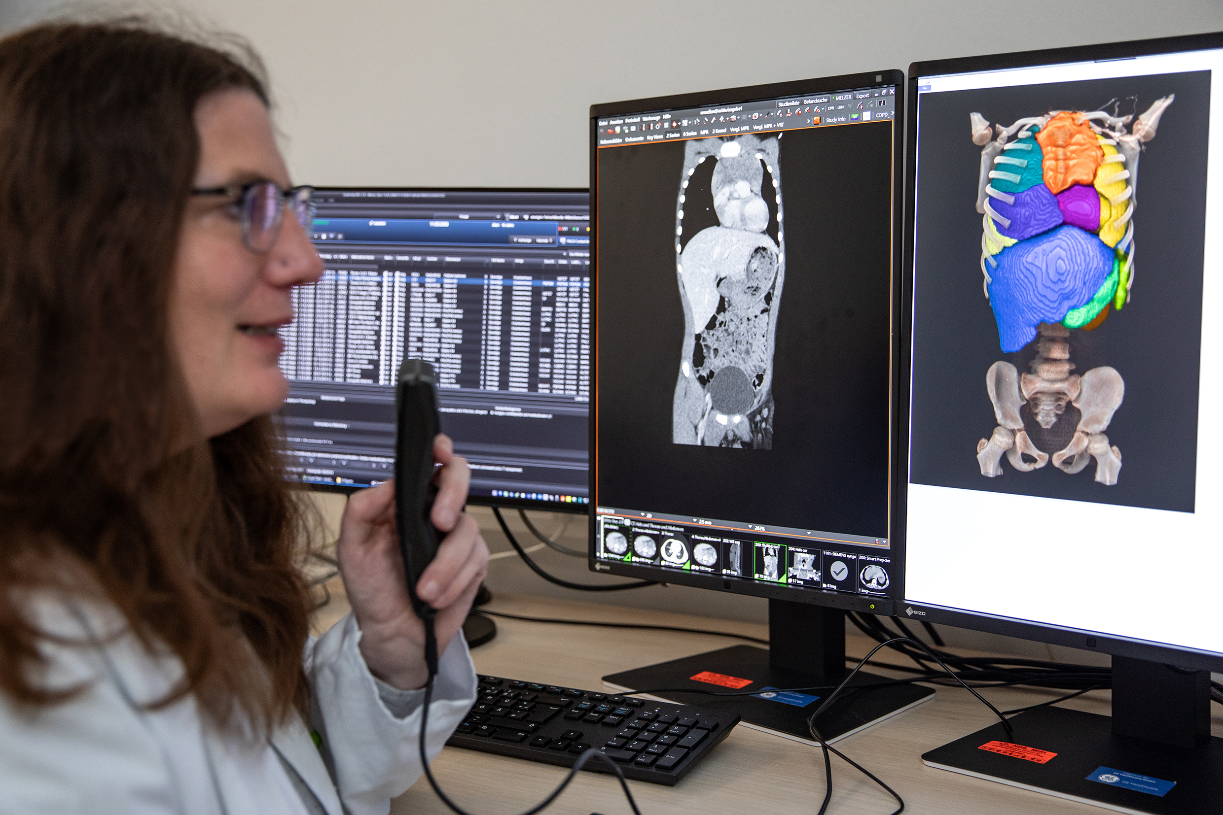
The RACOON-RESCUE project, led by the MHH, aims to automatically analyse image data from CT and MRI and find new image-based features in order to be able to more reliably assess the stage of disease in children and adolescents with non-Hodgkin's lymphoma.

In the RACOON-RESCUE project, Professor Dr Diane Renz wants to use radiological image data from CT and MRI to improve the assessment of non-Hodgkin's lymphomas in children and adolescents. Copyright: Karin Kaiser/MHH
Non-Hodgkin's lymphomas (NHL) are malignant diseases of the lymphatic tissue and are the fourth most common form of cancer in children and adolescents. More than 30 subgroups are known. The long-term survival rate is between 70 and 90 per cent. However, in case of a relapse, the overall prognosis for recovery is rather poor. Diagnosis is usually made using invasive methods - i.e. direct interventions in the body - such as a biopsy of the affected tissue or lumbar and bone marrow punctures. Non-invasive radiological diagnostics using imaging is a key component in investigating how far the lymphoma has spread in the body. However, there is still no standardised and automated image analysis that can be used to reliably and comparably determine the current stage and further progression of the disease.
The RACOON-RESCUE project now aims to change this. Under the leadership of Professor Dr Diane Renz, Head of Paediatric Radiology at the Institute of Diagnostic and Interventional Radiology at Hannover Medical School (MHH), researchers from the fields of paediatric radiology and paediatric oncology from 38 university hospitals want to jointly record and evaluate existing image data in a structured manner in order to better determine the stage of the disease, how patients are responding to treatment and what follow-up care is being considered. They also want to develop new image-based features. The research project is part of the Network of University Medicine (NUM) and is being funded by the Federal Ministry of Education and Research with a total of around one million euros over 18 months. Around 460,000 euros of this will go to the MHH.
Filtering information from radiological images
NHL is caused by changes in the genetic material of lymphocytes, which are important for immune defence. The lymph nodes are usually affected, but bone marrow, lungs, liver and spleen can also be affected in advanced stages of the disease. The disease typically begins with a painless swelling of the lymph nodes, which can also cause abdominal pain or breathing difficulties as the disease progresses, as well as fever, weight loss, night sweats and fatigue. Chemotherapy is usually used to treat NHL. In some cases, a stem cell transplant is necessary. ‘To assess the response, we want to rely even more on radiological examinations, which are less stressful for the children and adolescents affected,’ says Professor Renz. However, a method must first be developed to filter and process the flood of information from radiological images such as computed tomography (CT) and magnetic resonance imaging (MRI) in such a way that they can be more clearly assigned to a specific lymphoma stage.
Combining clinical and radiological data
‘Up to now, CT and MRI have not been used to determine a reference standard that radiologists can use as a guide,’ explains the head of the project. ‘This means that a large number of possible available indications of the tumour stage and the risk of recurrence unfortunately remain unused.’ A key aim of RACOON-RESCUE is therefore to develop a standardised and automated image analysis. The project is not only based on radiological image data that has already been recorded as part of routine clinical practice. The researchers are also utilising the ‘NHL-Berlin-Frankfurt-Münster Registry’, which has been collecting clinical data from children and adolescents of all NHL subgroups in Germany since 2012.
‘This gives us a new and unique data set that we want to analyse automatically with the help of artificial intelligence in order to improve predictions about the success of the therapy and predict the individual risk of relapse,’ explains Professor Renz. Image-based features such as the size, type and shape of the lymph nodes will also be included and help to train the machine prediction model. ‘Ultimately, we want to use this to develop a digital diagnostic mask to determine the respective tumour stage directly from the CT and MRI images, assess the response to therapy and thus also improve tumour aftercare.’ In the future, automated image evaluation should make it possible to use radiological diagnostics more comprehensively, reduce the use of invasive examinations such as tissue or fluid sampling and better classify previously unclear findings. ‘This will also make it possible to find out which patients have a high risk of relapse after treatment and the treatment concept can be customised.’
RACOON-RESCUE and the University Medicine Network
The RACOON-RESCUE project is coordinated by the Institute of Diagnostic and Interventional Radiology and the Department of Paediatric Haematology and Oncology at the MHH in cooperation with the Department of Paediatric Haematology and Oncology at the University Medical Center Hamburg-Eppendorf. All 38 German university hospitals affiliated to the Network of University Medicine (NUM), the German Cancer Research Centre in Heidelberg and three technical partners are involved.
The Network of University Medicine (NUM) consists of a central coordination office and the decentralised local staff units (LokS) of the 36 participating university hospitals in Germany. The LokS support cooperation in the NUM's cross-site infrastructures and research projects. They are the point of contact for NUM project participants, promote cooperation between researchers at national level and are informed about all ongoing NUM projects. In the first funding phase (2020-2021), MHH researchers took part in eight collaborative projects, in the second funding phase (2022-2025) in 19 projects. The application period for the third funding phase starts this summer. Contact: num-loks@mh-hannover.de.
Text: Kirsten Pötzke