Imaging technique developed at MHH enables regional imaging of lung function without radiation exposure and breathing stops.
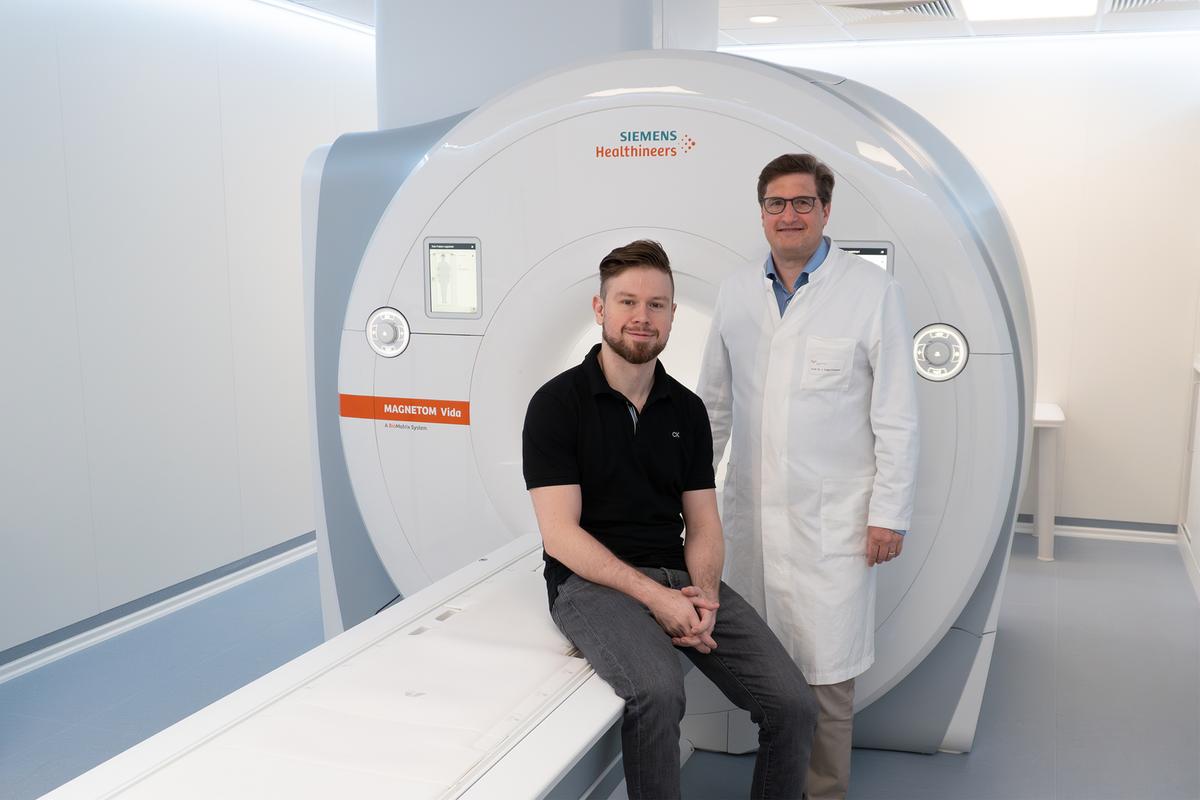
Developed the PREFUL method for regional lung imaging in MRI: Dr. Andreas Voskrebenzev, physicist (left) and Professor Dr. Jens Vogel-Claussen (right), Senior Consultant at the Institute of Diagnostic and Interventional Radiology at the MHH. Copyright: Dr. Andreas Voskrebenzev/MHH
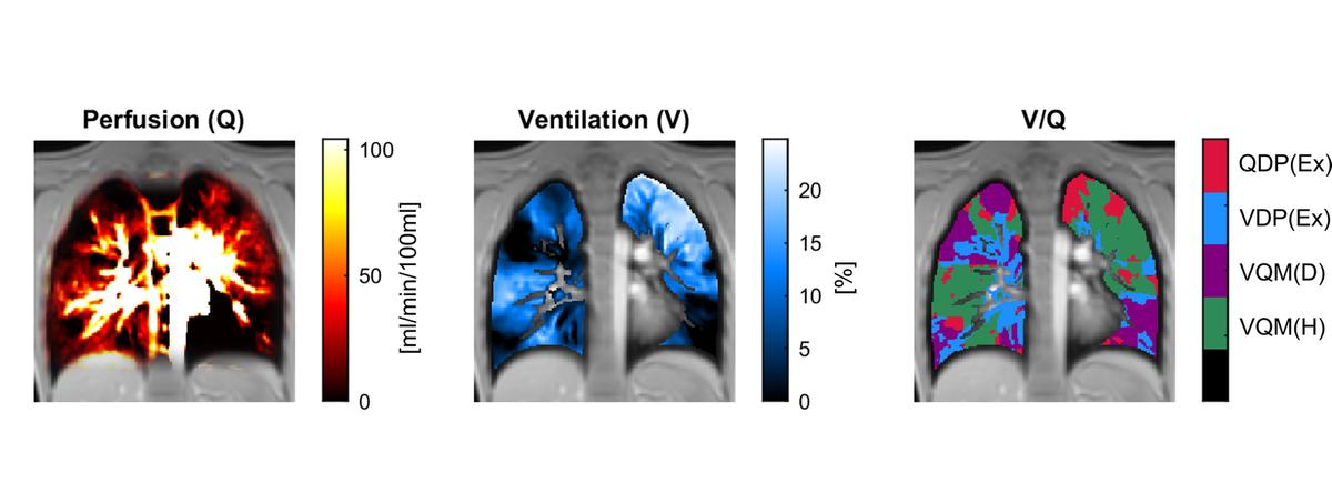
Three exemplary PREFUL images of a cystic fibrosis patient show information about blood flow (perfusion) and ventilation of the lungs. Both the individual maps (left and center, dark shades) and the combined V/Q map (right) clearly show that there are many areas with impaired lung function (red, blue, purple) outside of normal lung function (green). Copyright: Dr. Andreas Voskrebenzev/MHH
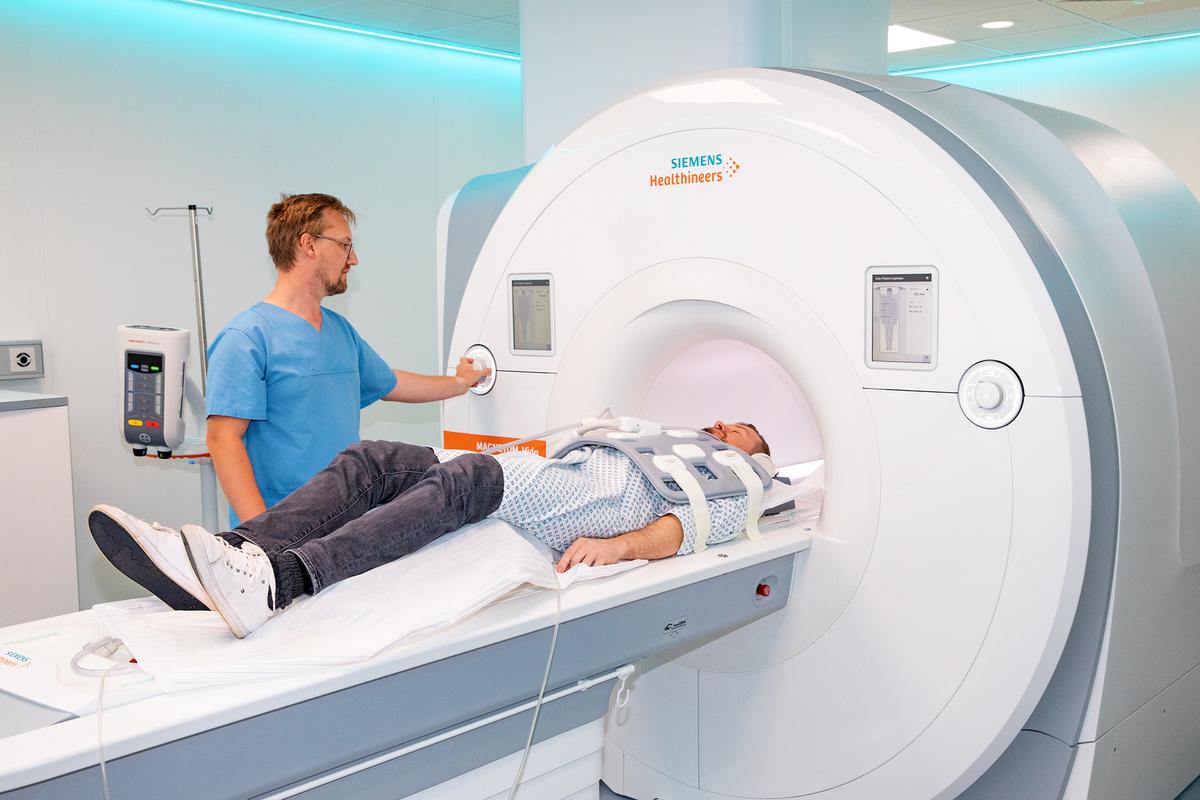
Uses the PREFUL method for regional lung imaging in MRI: MHH radiologist Dr. Till Kaireit. Copyright: Karin Kaiser/MHH
Recognizing and assessing lung diseases is a medical challenge. Conventional computed tomography (CT) is well suited to depicting the structure of the lungs in high resolution. However, it provides little information about lung function and also exposes patients to radiation. The lung function test is well established, but only provides values for the entire lung, i.e. it only tells us whether the lung is diseased, but not exactly where. An alternative is imaging using magnetic resonance imaging (MRI), which works without radioactive radiation. However, the lungs are a difficult organ for MRI due to their high air content, as only tissue containing hydrogen can be depicted well in the MR image. It is also important that patients hold their breath repeatedly during conventional MRI lung imaging.
Specialist journal publishes article and detailed video instructions
In order to make lung MRI more patient-friendly and generally accessible, researchers led by Professor Dr. Jens Vogel-Claussen, Senior Consultant at the Institute of Diagnostic and Interventional Radiology at Hannover Medical School (MHH) and the German Center for Lung Research in Hannover (DZL-BREATH), have developed the so-called PREFUL method. This imaging technique allows the ventilation and blood flow of the lungs to be visualized regionally with a conventional MRI device in high temporal resolution and does not require any contrast agent, specially developed MRI software (sequence) or breathing stops. To ensure that PREFUL MRI benefits as many patients as possible, the researchers have now published the entire examination procedure with all the calculations required for image evaluation in the “Journal of Visualized Experiments”. The special thing about this is that the electronic journal not only publishes the detailed instructions as a specialist article, but also makes them available online as a detailed video recording.
No additional devices or personnel required
PREFUL stands for “phase-resolved functional lung”. This MRI method records signal changes in the lungs over the entire respiratory and cardiac cycle. It therefore not only measures how the density of the lung tissue changes during inhalation and exhalation, but also records the changes when the heart pumps blood into the body and thus through the imaging plane. “This now enables us to quantify regional lung function in early-stage lung diseases, improve therapy monitoring and also predict disease progression after lung transplantation, for example,” explains Professor Vogel-Claussen.
“PREFUL MRI has many advantages,” says MRI physicist Dr. Andreas Voskrebenzev, who played a key role in developing the imaging technology. “It provides spatially resolved functional information on exactly where the problem is in the lungs, makes the examination less stressful for patients and requires no additional technical equipment or medical staff other than a standard MRI machine.” This method, which is already well established experimentally, is therefore particularly useful for specialist radiology practices and clinics that are interested in so-called exploratory imaging markers. These are measurements of lung function that are not yet clinically established, but are used in research and are suitable for monitoring sensitive patients, for example.
PREFUL-MRI tested in numerous studies
Around three to five images per second are taken during the examination. Each of these is assigned to a specific respiratory and cardiac phase. “We can re-sort the images after the MRI examination in order to combine several images of a very specific respiratory phase and a very specific cardiac phase and thus achieve a greater temporal resolution, i.e. obtain more information,” explains Dr. Voskrebenzev. The images can then be converted into biomarkers and can, for example, indicate the blood flow in the lungs as a specific value in milliliters per minute. The researchers have tested the method in numerous studies using various clinical pictures and different age groups. For example, they were able to show that the respiratory function of COPD patients improved significantly after inhalation treatment. “This was directly visible in the lungs,” says the scientist.
Better prediction of complications after transplantation
PREFUL-MRI has also proven its worth in patients with cystic fibrosis, pulmonary hypertension and COVID-19 in order to assess the diseases and monitor changes in treatment after therapy. PREFUL-MRI can also be used to better predict chronic complications after lung transplantation. With their publication, the researchers are now issuing a kind of manual with precise instructions, possible sources of error and suggested solutions for their PREFUL method. Their aim is to make the technique accessible to all interested radiologists and to further promote the spread of PREFUL MRI.
Those who do not want to work their way through the methodological instructions in order to use the PREFUL method in their own practice or clinic can also obtain the software as a ready-to-use app. The start-up “BioVisioneers”, founded by the MHH researchers, provides interested parties with the PREFUL app for immediate use for a fee.
Text: Kirsten Pötzke