Gastroenterology puts new X-ray system into operation

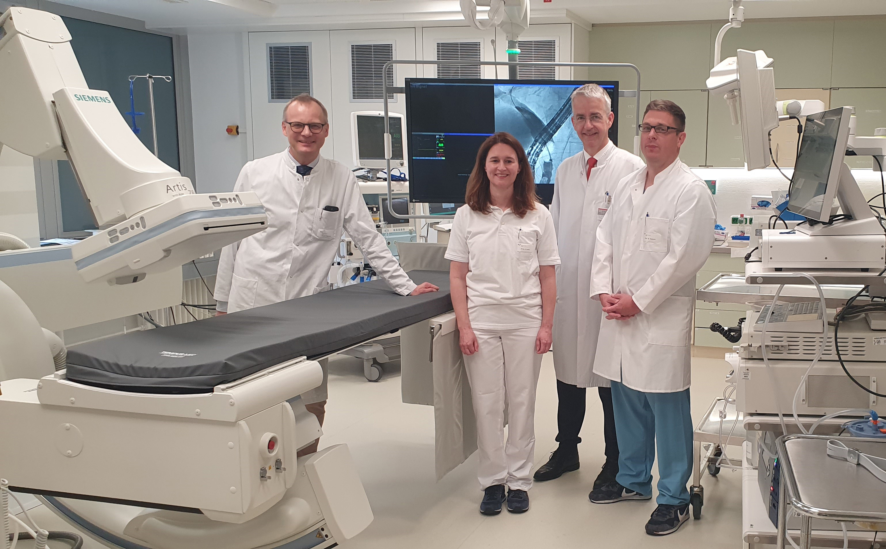
Showing off the new X-ray system: Clinic Director Professor Dr. Heiner Wedemeyer, PD Dr. Henrike Lenzen, MHH Vice President Professor Dr. Frank Lammert and Professor Dr. Benjamin Heidrich. Copyright: Inga Budde / MHH.
Great joy at the Clinic for Gastroenterology, Hepatology, Infectiology and Endocrinology: a new X-ray system has now been put into operation. The state-of-the-art device called Artis zee is characterised by many technical innovations that benefit both patients and staff. "The system offers us even better options for diagnosing and treating diseases of the gastrointestinal tract, thereby strengthening patient care at the highest level," says Clinic Director Professor Dr Heiner Wedemeyer.
The new device is used in endoscopy. An endoscopy is also known as optical imaging. Probes fitted with tiny cameras are inserted into the body to assess the condition of organs and, if necessary, to carry out treatment directly. Artis zee is equipped with innovative imaging technology, allowing experts to recognise details and display a wealth of information. The system delivers high image quality with less radiation - this is a great advantage for patients, but also for the endoscopy team. After all, staff are challenged every day to protect themselves from scattered radiation.
Recognise and treat immediately
"We will use the system for examinations of the entire gastrointestinal tract," explains Professor Dr Benjamin Heidrich, Head of the Endoscopy Department. An endoscopy normally takes one to two hours. Professor Heidrich therefore expects to be able to carry out around four examinations per day on the new device. One of the most frequent examinations that he and his team carry out is endoscopic retrograde cholangiopancreatography (ERCP).This involves visualising the bile ducts and the pancreatic duct system.
"If we detect gallstones or constrictions, for example, we can treat them immediately using imaging," explains Heidrich, who took over the management of the department from PD Dr Henrike Lenzen at the beginning of July 2020. Dr Lenzen had been the medical coordinator for the conversion measures in recent years.
Another typical examination is percutaneous transhepatic cholangiodrainage (PTCD). In this procedure, a drain is inserted into the bile ducts of the liver in order to dissolve a blockage of bile.
The installation of the new X-ray system was not an easy task, as it was carried out during ongoing operations. "But thanks to good coordination with the endoscopy team, everything went very smoothly," says Kerstin Zimmermann from MHH construction management. As part of the remodelling work, all the supply lines, the entire electrical system and the floor were renewed.
Text: Tina Götting